
In this case, the uppermost fragment of the ossicle was of abnormal low signal intensity on T1-weighted MR images with corresponding high signal intensity on T2-weighted and STIR images, indicating bone marrow oedema due to post-traumatic fracture. The latter are commonly caused by inversion injuries in the clinical setting of ankle sprains. Furthermore, the ossicle may cause limitation of the range of motion of the ankle joint resembling avulsion fractures of the lateral malleolus. Mechanical irritation or joint instability may cause pain and recurrent ankle sprains. It has been postulated that symptoms are associated with disruption to the fibrous or cartilaginous attachments of the ossicle resulting in a fracture, fibrous union, or pseudarthrosis.

Ossicles may also be connected to the fibres of the posterior talofibular ligament. Arthroscopic and operative findings have shown that os subfibulare is embedded partially or completely within the fibres of the anterior talofibular ligament, with some parts exposed in the talofibular joint or covered by a synovial membrane. The os subfibulare most commonly remains asymptomatic, however, it may cause pain, sustain or simulate a fracture, or it may even precipitate arthrosis in response to overuse and trauma. The os subfibulare is usually round, oval, or comma-shaped. Such ossicles rarely persist beyond skeletal maturation with a reported prevalence of 1- 2.1%. It appears toward the end of the first year of life and fuses with the metaphysis between the ages of 15 and 17 years. The ossicle is located under the tip of the lateral malleolus. If you need medical advice, please contact a health care professional.The os subfibulare is a normal anatomic variant that represents either an unfused accessory ossification centre or a supernumerary bone. We cannot respond to patients or patient advocates requesting advice on issues related to medical conditions.
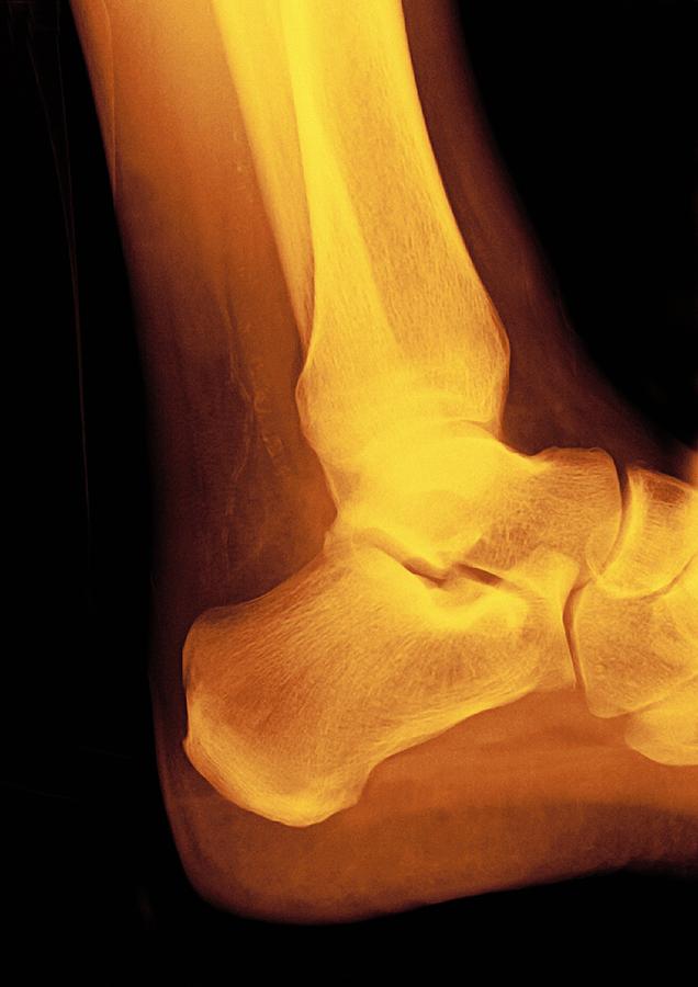
Normal ankle xray professional#
The Guidelines are not intended as a substitute for the advice or professional judgment of a health care professional, nor are they intended to be the only approach to the management of clinical problems. The Guidelines are intended to give an understanding of a clinical problem and outline one or more preferred approaches to the investigation and management of the problem. The principles of the Guidelines and Protocols Advisory Committee are to:ĭisclaimer The Clinical Practice Guidelines (the "Guidelines") have been developed by the Guidelines and Protocols Advisory Committee on behalf of the Medical Services Commission. This guideline was developed by the Guidelines and Protocols Advisory Committee, approved by the British Columbia Medical Association and adopted by the Medical Services Commission. This guideline is based on scientific evidence current as of the Effective Date.
Normal ankle xray trial#
Multicentre trial to introduce the Ottawa Ankle Rules for use of radiography in acute ankle injuries. Managing ankle sprains in primary care: what is best practice? A systematic review of the last 10 years of evidence. Implementation of the Ottawa Ankle Rules. Stiell I, McKnight RD, Greenburg GH, et al.Diagnosis and treatment of acute ankle injuries: development of an evidence-based algorithm. Clinical usefulness of the Ottawa Ankle Rules for detecting fractures of the ankle and midfoot. Cost-effectiveness analysis of the Ottawa Ankle Rules. Anis AH, Stiell IG, Stewart DG, Laupacis A.


 0 kommentar(er)
